
Figure 4. MC distribution in tissues after oxazolone-dermatitis and OVA-induced asthma in i.d. reconstituted KitW-sh/W-sh mice.
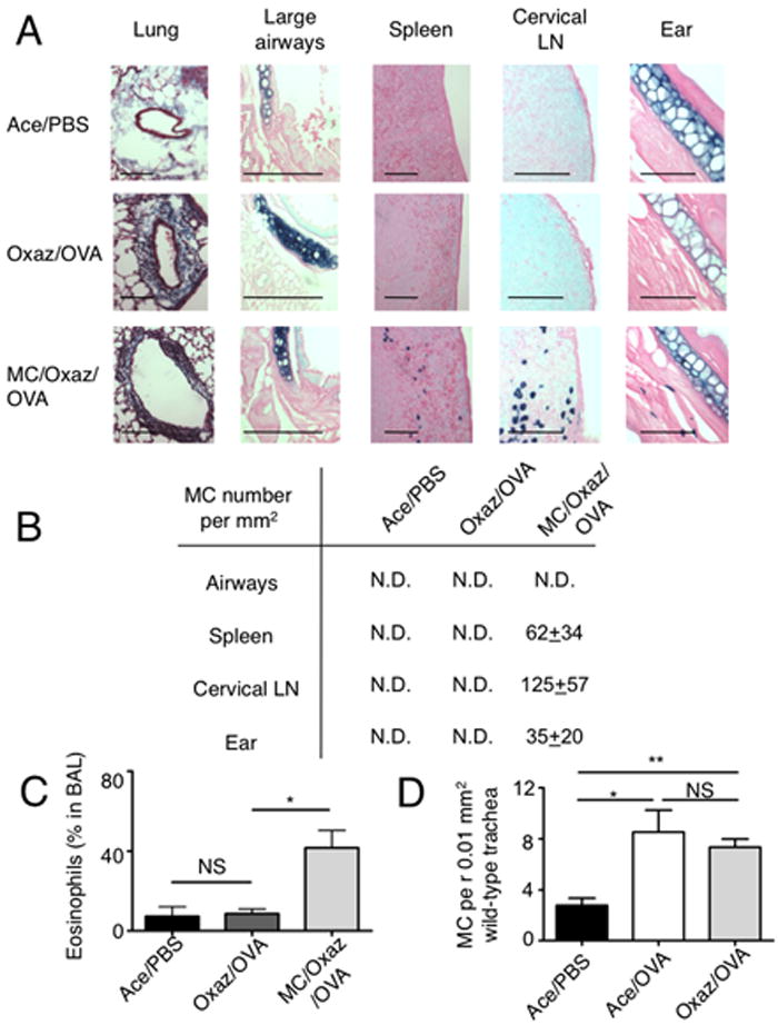
(A) Tissues were harvested from the indicated three groups of mice and embedded in paraffin after the OVA challenge. Lung samples were stained with Masson’s trichrome to demonstrate inflammatory infiltrate within peri-bronchiolar collagen. Large airways, spleens, cervical LN and ear tissues were stained for MCs, with toluidine-blue. Bars, 100 μm. (B) Quantitation of MCs in tissues of mice with asthma was done under a light microscope and is presented as mean±sem per mm2. N.D., not detected. For comparison, the basal MC numbers in unchallenged i.d. reconstituted mice were: airways, N.D; spleen, 4.5±1.5; cervical LN, 218±59, ear, 27±6 (n=5). (C) : Quantitation of eosinophils in bronchoalveolar lavage (BAL) fluid of mice in (A, B) (n=3). *, p=0.02. (D) MCs in trachea tissue of wild type Balb/c mice, enumerated following toluidine blue staining under a light microscope (n=5). *, p=0.016; **, p=0.0011.