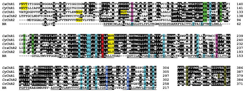
Figure 2.
Partial alignment of Chlamydomonas channelopsin and BR sequences. Black background indicates conserved identical residues. Turquoise background, positions of the residues that form the retinal-binding pocket in BR. Green background, conserved Glu residues in the predicted second helix. Magenta background, molecular determinants that differentiate CrChR1/VcChR1 from CrChR2/VcChR2. Red background, residues in the position of the proton donor in BR. Blue background, residues in the positions of Glu194 and Glu204 in BR. Yellow background, predicted glycosylation sites. Olive background, conserved residues known to be phosphorylated in CrChR1 or CrChR2. Underlined characters show the regions that form transmembrane helices in BR.

