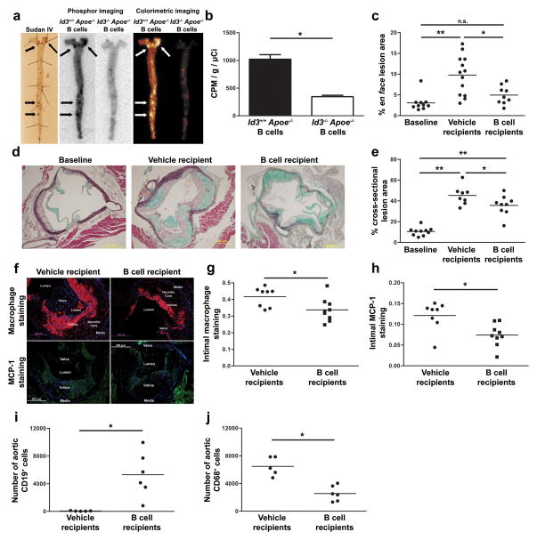
Figure 6. Adoptively transferred B cells traffic to regions of atherosclerosis, decrease plaque macrophage content and attenuate atherosclerosis progression.
a, Sudan IV staining and ex vivo phosphor imaging of aorta from μMT Apoe−/− mice fed eight weeks of Western diet and injected with 2 × 10⁷ In-111 radiolabeled Apoe−/− or Id3−/−Apoe−/−B cells. b, Quantification (n=4) of counts adjusted for injected radioactivity and aortic weight. *:p=0.0003. c, Quantification of en face lesion area of μMT Apoe−/− mice fed 8 weeks of a Western diet (baseline, n=10) or injected with vehicle control (n=13) or 45 × 106 Apoe−/− B cells (n=9) followed by an additional 8 weeks of Western diet *:p=0.011, **:p=0.0005, n.s. = not significant. d, Representative images and e, quantitation of MOVAT stained cross-sections at the aortic cusp *:p<0.05, **:p<0.001. f, Representative images and quantitation of g, macrophage and h, MCP-1 content in μMT Apoe−/− mice with 8 weeks of prior Western diet feeding that received injection of vehicle control or Apoe−/− B cells and an additional 8 weeks of Western diet feeding. Aortic cross sections (4 per animal at 60 μm intervals) were stained using DAPI (blue) and either a Mac-2 monoclonal antibody (red) or a MCP-1 polyclonal antibody (green) *:p=0.024 and 0.007 respectively. Quantitation of aortic i, B cell (CD19+ CD45+) and j, macrophage (CD68+ CD45+) content determined by flow cytometry 72 hours following adoptive transfer of either PBS vehicle injection or 60 × 106 Id3+/+ Apoe−/− B cells to chow-fed μMT Apoe−/− mice. *:p=0.006 and 0.0005 respectively.

