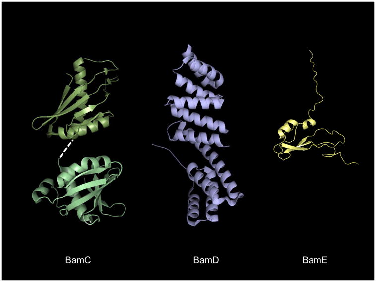
Figure 6.
The solution structures of E. coli BamCDE. The BamD (PDB ID: 2YHC) and BamE (PDB ID: 2KM7) structures are oriented with the N-termini pointing toward the top of the page. The structurally homologous helix grip domains of BamC (PDB ID: 2LAE, 2LAF) are shown on the left, with the extreme C-terminal domain at bottom. A dashed white line indicates the unresolved helix linking the two domains.

