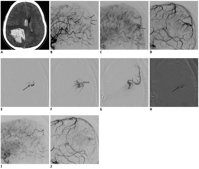
Fig. 1.
18-year-old male with atypical developmental venous anomaly.
A. Initial precontrast brain CT image showing large intracerebral hemorrhage in right parietal lobe and intraventricular hemorrhage in bilateral lateral ventricles. B. Right internal carotid angiogram of lateral image showing arterial pedicle (arrows) from pericallosal branch of right anterior cerebral artery. C. Late arterial phase angiogram showing early venous drainage (arrow). D. Developmental venous anomaly visualized in right parietal lobe in venous phase. E-G. Serial microangiogram clearly demonstrating arterial pedicle (arrowheads), site of arteriovenous fistula (large arrow) and early venous drainage (small arrows). Venous drainage from arteriovenous fistula shares venous channel of developmental venous anomaly showed in D and tip of microcatheter (open arrow) is also seen. H. Cast of NBCA-Lipiodol is located in distal arterial pedicle (arrowhead), arteriovenous fistula (large arrow), and venous channel just distal to arteriovenous fistula (small arrow). I, J. Late arterial phase lateral projection image (I) showing delayed flow in pedicle along with no arteriovenous shunt in post-embolization angiogram. Developmental venous anomaly still persists in venous phase (J) after embolization.

