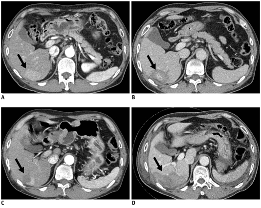
Fig. 2.
63-year-old man with 4.3-cm-diameter hepatocellular carcinoma treated by switching monopolar radiofrequency ablation.
A. Pre-ablation CT scan during arterial phase showing hyperenhancing 4.3-cm-diameter hepatocellular carcinoma nodule (arrow). B. Immediate post-ablation CT scan during portal phase showing no definite residual enhancing tumor (arrow). C. Arterial phase nine-month follow-up CT scan showing local tumor progression (arrow) on superior side of radiofrequency ablation defect. D. Follow-up CT scan obtained immediately after second radiofrequency ablation using two single electrodes in switching monopolar mode showing complete ablation of recurrent hepatocellular carcinoma (arrow).

