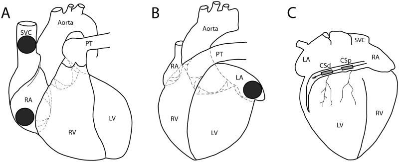
Figure 1.
Anatomic Positions of Defibrillation Electrodes. Schematic of a canine heart with locations of electrodes from which defibrillation therapies were delivered depicted. A: right anterior oblique (RAO) view showing superior vena cava and right atrial appendage disc electrodes. B: left anterior oblique (LAO) view showing left atrial appendage disc electrode. C: posteroanterior (PA) view showing distal and proximal coronary sinus coils. SVC, superior vena cava; RA, right atrium; PT, pulmonary trunk; RV, right ventricle; LV, left ventricle; CSp, proximal coronary sinus; CSd, distal coronary sinus.

