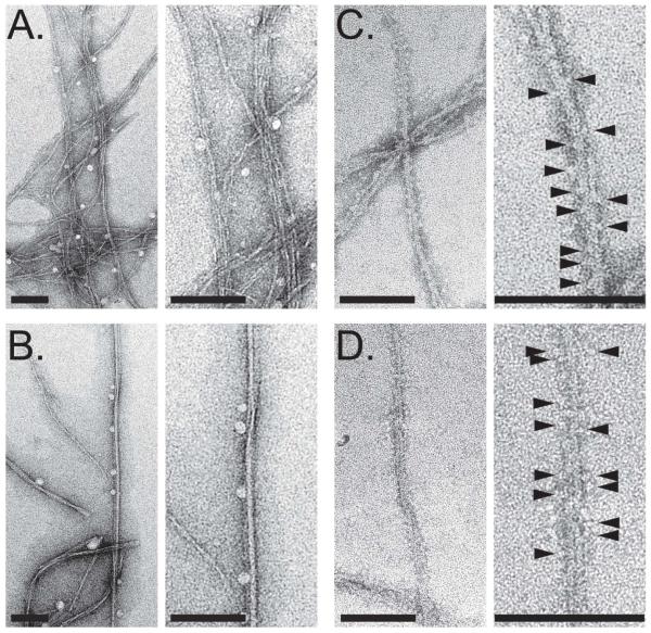
Figure 3. Transmission electron micrographs of 1541 nanofibrils with and without procaspase-3.
(A) Negatively stained 1541 at 25°C shows bundles of very thin and flexible fibrils. (B) In contrast, 1541 at 37°C consists mainly of larger, less flexible fibrils. (C) 1541 fibrils at 25°C decorated with procaspase-3; and (D) 1541 fibrils at 37°C decorated with procaspase-3. In (C) and (D) the surface of the fibrils appears decorated with procaspase-3 molecules (arrow heads). Magnification bars = 100 nm.

