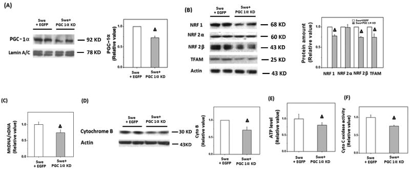
Figure 5.
Transfection with miR RNAi of PGC-1α exacerbated the impaired mitochondrial biogenesis in APPswe cells. A) Representative immunoblotting and quantification analysis showed that the expression of PGC-1α significantly reduced after transient transfection with miR RNAi of PGC-1α. B) Representative immunoblotting and quantification analysis revealed that the levels of NRF 1, NRF 2 and TFAM were significantly reduced after transient transfection (*p<0.05, Δp<0.01, Student's t test). C) Real time PCR revealed that mtDNA was significantly reduced after transient transfection with miR RNAi of PGC-1α for 48 h compared with EGFP control cells (Δp<0.01, Student's t test). D) Representative immunoblotting and quantification analysis revealed cytochrome B was significantly reduced (Δp<0.01, Student's t test) after cells was transfected with miR RNAi of PGC-1α for 48 h. E) Firefly luciferase assay showed that ATP level displayed a significant decrease after transient transfection with miR RNAi of PGC-1α for 48 h in APPswe cells (Δp<0.01, Student's t test). F) Cytochrome C oxidase activity was significantly decreased after transfection with miR RNAi of PGC-1α in APPswe cells (Δp<0.01, Student's t test).

