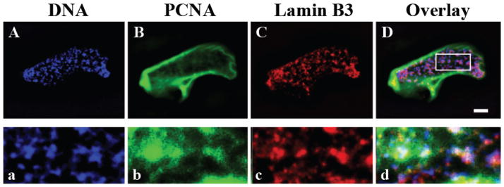
Figure 3.
Lamin B3 is closely associated with PCNA and chromatin in in vitro assembled nuclei. Localization of Xenopus lamin B3 (B, b; green) and PCNA (C, c; red) in a nucleus assembled in a Xenopus egg interphase extract for 130 min (A–D). DNA is stained with Hoechst dye (A, a; blue). The area in the box in D is enlarged (3.5×) to show the partial overlap between DNA, lamin B3 and PCNA (a–d). Brightness and contrast are enhanced in b compared to B for better visualization of the internal lamin B3 structures. Scale bar, 5 μM.

