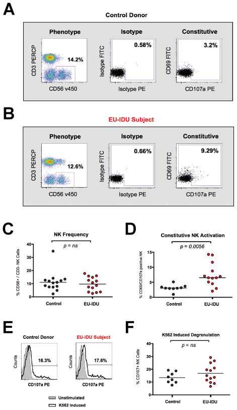
Figure 1. NK cells from EU-IDU subjects exhibit increased activation and maintain normal function.
(A–B) PBMCs from one representative control donor (A) and one representative EU-IDU subject (B) were stained with fluorescently conjugated antibodies to NK phenotypic and functional markers (CD3, CD56, CD69, CD107a). NK cells were gated as CD56+/CD3− lymphocytes (first panel) and the frequency of constitutive CD107a (x-axis) and CD69 (y-axis) staining is shown compared to isotype control. (C–D) Composite graph of the frequency (C) and constitutive CD69/CD107a staining (D) on CD56+/CD3− NK cells from control uninfected donors and EU-IDU subjects as shown in panel A and B. Statistical analyses were performed using a non-parametric Mann-Whitney T-test with a two-tailed p-value. (E) PBMCs from one representative control donor and one representative EU-IDU subject were incubated for 3 hours with a fluorescently conjugated mAb to CD107a in the presence or absence of MHC-devoid K562 cells at a 10:1 effector to target cell ratio. PBMCs were then stained with fluorescently conjugated antibodies to NK phenotypic markers (CD56/CD3) and the percentage of CD56+/CD3− gated NK cells staining positive for CD107a is shown on unstimulated (light gray, shaded histogram) and K562 stimulated (bold, open histogram) PBMCs. (F) Composite graph of K562-induced CD107a staining on CD56+/CD3− gated NK cells from control uninfected donors and EU-IDU subjects as shown in panel E.

