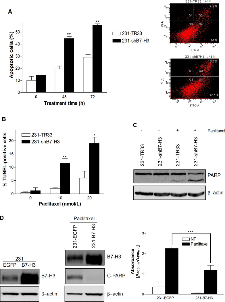
Figure 3.
B7-H3 silencing sensitizes breast cancer cells to paclitaxel-induced apoptosis. A, The percentage of Annexin V- FITC stained cells was increased in B7-H3 knockdown population. 231-TR33 and 231-shB7-H3 cells were treated with 20 nmol/L paclitaxel for 0, 48, and 72 h, respectively, and apoptosis was examined by Annexin V-FITC staining and flow cytometry. Left: The percentage of apoptotic cells; Right: Flow cytometry diagram at 48 h. B, The percentage of TUNEL-positive cells was increased in B7-H3 knockdown population. Cells were treated with 0, 10 and 20 nmol/L paclitaxel for 72 h, and apoptosis was examined by TUNEL assay and flow cytometry. C, PARP cleavage was increased in B7-H3 knockdown cells, as detected by Western blotting following treatment with 20 nmol/L paclitaxel for 72 h. β-actin was used as a loading control. D, B7-H3 overexpressing MDA-MB-231 cell varaiants were more resistance to pacilitaxel-induced apoptosis. Left: B7-H3 overexpressing 231-B7-H3 cells expressed higher level of B7-H3, compared to the 231-EGFP control cells as detected by Western blot analysis; Middle: B7-H3 overexpressing cells showed decreased level of cleaved-PARP compared to the control cells upon treatment with 20nmol/L paclitaxel for 72 h; Right: apoptosis-specific ELISA detection revealed that the level of cytoplasmic histone-associated DNA fragments in paclitaxel treated 231-B7H3 cells was significantly lower than in control 231-EGFP cells. Bars, S.E.; * p< 0.05; ** p< 0.01; *** p < 0.001.

