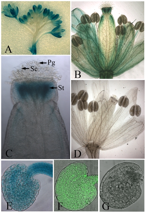
Figure 1. Expression of the Prosiz1::GUS–GFP gene.
(A) Expression of Prosiz1::GUS–GFP in the whole inflorescence. A GUS signal was detected in most of the flowers, except the latest ones, while the strongest GUS signal was found in sepals. The inflorescence in (A) was stained with 1 mM 5-bromo-4-chloro-3-indolyl-b-glucuronic acid (X-Gluc) for 12 h. (B) GUS signal in a flower after pollination. The style of the pistil was stained strongly by X-Gluc, and the upper part of the carpel and the stem of the stamen were also stained by X-Gluc. No GUS signal was seen in the anthers. (C) GUS signal in reproductive organs at the end of pollination. The style was strongly stained by X-Gluc, while no GUS signal was seen in the stigmatic cells or pollen. (D) No GUS signal was detected in the whole flower in the wild-type plants after staining with 1 mM X-Gluc for 12 h. (E) Expression of Prosiz1::GUS–GFP could be detected in all cells within the ovule before fertilization after staining with 1 mM X-Gluc for 8 h. (F) GFP fluorescence of Prosiz1::GUS–GFP can be seen in all cells of the ovules. (G) Wild-type ovule control. No fluorescence was detected in the wild-type ovule under LSCM. Pg, pollen grain; Sc, stigmatic cell; St, style.

