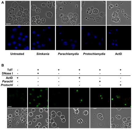
Figure 3. Parachlamydiaceae-induced cell death in S2 cells is accompanied by apoptosis-like morphological and nuclear changes and DNA fragmentation.
Panel (A) illustrates morphological and nuclear changes that were observed in S2 cells in response to infection with Parachlamydiaceae, but not Simkania. Insect cells were infected with S. negevensis, Pa. acanthamoebae, or P. amoebophila (MOI 2.5). At 7 h p.i. DNA was stained with DAPI (blue). Untreated cells and cells treated with the apoptosis inducer ActD (7 h) are shown for comparison. The bar corresponds to 10 µm. In (B) the detection of DNA fragmentation by TUNEL staining in S2 cells infected with Parachlamydiaceae is shown. S2 cells were either left untreated, incubated with ActD (10 h), or infected with Pa. acanthamoebae or P. amoebophila (MOI 2.5, 10 h). Bacteria were detected by immunostaining using antibodies raised against purified bacteria (red). TUNEL-positive nuclei are shown in green. Two additional controls were included, a negative control where TdT was omitted from the TUNEL reaction mixture and a positive control where cells were preincubated with DNase I to experimentally introduce DNA double strand breaks in all (also non-apoptotic) cells. Note that apart from this control, TUNEL-positive cells typically display other characteristic features of apoptotic cells, such as condensed and fragmented nuclei and formation of apoptotic bodies. After infection, TUNEL-positive cells were also frequently associated with bacteria. The bar indicates 10 µm.

