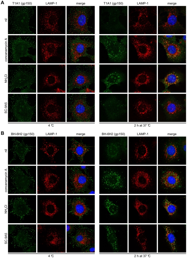
Figure 7. Gp150 undergoes pre-fusion antigenic changes.
Infections, drug treatments and antibody treatments were as for Figure 3. In (A) the cells were stained with the gp150-specific IgG2a BN-3A4, in (B) with the gp150-specific IgG2a T1A1 (green), and in (C) with the gp150-specific IgG2b BH-6H2. The cells were also stained for LAMP-1 (red) and with DAPI (blue). Treating cells with DMSO alone had no effect (not shown).

