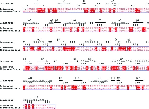
Figure 5.
Amino-acid sequence alignment of Mtb HisC2 and its counterpart from L. innocua. The secondary-structural elements (helices represented by coils, β-strands by arrows and turns by letter Ts) corresponding to histidinol phosphate aminotransferase from L. innocua are shown. Conserved residues are highlighted with a red background. The sequence alignment was performed by the program MultAlin (Corpet, 1988 ▶) and the figure was generated using the program ESPript (Gouet et al., 1999 ▶).

