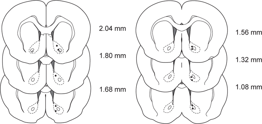
Figure 5.
Anatomical distribution of carbon fiber electrode recording sites in the NAc. Coronal sections show electrode tip locations for 11 recording locations. Numbers to the right indicate anterior posterior coordinates rostral to bregma. Coronal sections and coordinates adapted from stereotaxic atlas (37).

