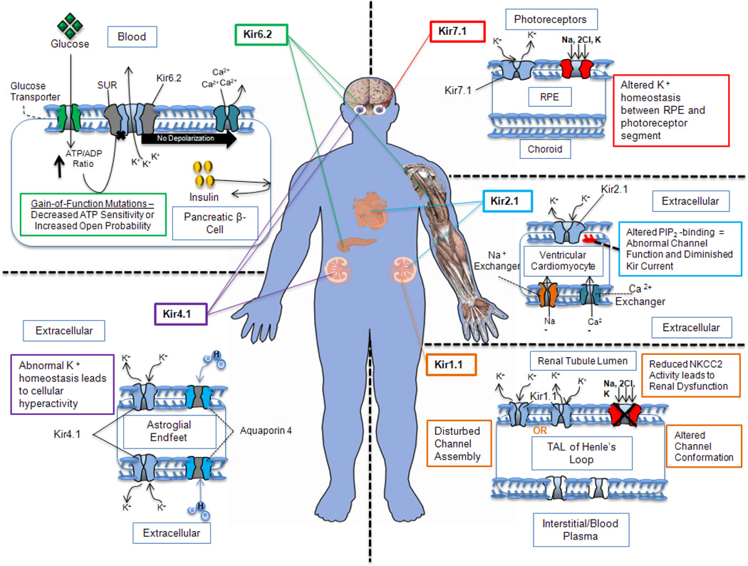
Figure 3. Tissue distribution of Kir channel subunits.
The tissue-specific distribution of the Kir channels suggests that they play an important role in ion homeostasis and disease. Kir channel subunits are indicated by light blue within the membrane structure. All other possible associated channels, transporters and regulatory molecules are also shown in the membrane that controls cellular physiology. Kir channels tissue distribution along with their respective physiopathology are color-coded (Kir1.1- orange; Kir2.1- blue; Kir4.1- purple; Kir6.2- green and Kir7.1- red). Abbreviations: Kir, inwardly rectifying potassium channel; SUR, regulatory suramine subunit; ATP, adenosine tri-phosphate; ADP, adenosine di-phosphate; RPE, retinal pigment epithelium; PIP2, phosphatidylinositol (4,5)-bisphosphate; TAL, thick ascending limb.

