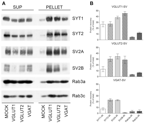
Figure 5.
Expression of SYT1, SYT2, SV2A, SV2B, and Rab3a and c in VGLUT1+, VGLUT2+, and VGAT+ SVs. (A) Glutamatergic and GABAergic SVs were immunoisolated from the LS1 fraction of rat cerebral cortex using beads coupled with either rabbit VGLUT1, VGLUT2, or VGAT antibodies. After immunoisolation, corresponding amounts of pellet and supernatant (SUP) fractions were subjected to immunoblotting with anti-SYT1 and -SYT2 antibodies, anti-SV2A/SV2B antibodies, or anti-Rab3a/Rab3c antibodies. (B) Quantification of the recovered immunoreactivities was carried out by densitometric scanning and interpolation of the data into a standard curve of rat brain LS1 fraction, and expressed as percent of the total input of LS1 added to the samples. The percentage of SYT1/SYT2, SV2A/SV2B, and Rab3a/Rab3c immunoreactivities (IR) detected in SVs immunoisolated with anti-VGLUT1 (VGLUT1–SV; upper left/right panel), anti-VGLUT2 (VGLUT2–SV; lower left/right panel), or anti-VGAT (VGAT SV; upper right/right panel) beads are shown as means (±SEM) of five independent experiments.

