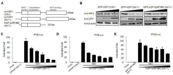
Figure 1.
Rotavirus NSP1 inhibits IFN-β promoter activation independent of IRF3 degradation. (A) Scheme of full-length (wt) NSP1 structures of rotavirus OSU and SA11 strains and C-truncated (ΔIRF3 binding domain) NSP1 mutant of rotavirus SA11. (B) Western blot analysis for degradation of IRF3 by OSU NSP1, SA11 NSP1 and NSP1ΔIRF3BD (SA11). 293FT cells were transfected with pCMV-IRF3, pEGFP-OSU NSP1 or pEGFP-SA11 NSP1 or pEGFP-NSP1ΔIRF3BD (SA11) plasmids and cell extracts were assayed 48 h post-transfection for the expression of IRF3. Immunoblots were probed with anti-IRF3 monoclonal antibody (top panel). β-actin was used as a loading control (bottom panel). (C, D, E) Inhibition of virus-induced IFN-β promoter activation by SA11 NSP1 (C), OSU NSP1 (D) or NSP1ΔIRF3BD (SA11) (E). 293FT cells were transfected with pGL3-IFN-β-Luc, pRL-SV40, and increasing amounts of SA11 NSP1, OSU NSP1 or NSP1ΔIRF3BD (SA11) expression plasmids. Cells were infected with Sendai virus for 24 h and assayed for luciferase activities. Data are expressed as folds of activation with standard deviations among triplicate samples.

