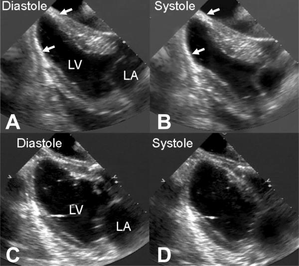
Figure 2.

Representative two-dimensional long-axis echocardiograms in diastole and systole in representative treatment (Panel A and B) and control (Panel C and D) hearts 8 weeks after infarction. Note the preservation of the normal elliptical left ventricular shape in the treated heart and the near spherical shape of the control heart. The white arrows identify the radio opaque filler material in the apical region of the treated heart.
