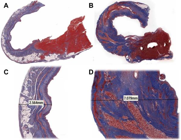
Figure 4.
Masson's trichrome staining in representative control (Panels A and C) and treatment (Panels B and D) animals 8 weeks after infarction. Panels A and B are 1× magnification and Panels B and D are 1.2×. Notice the exuberant collagen production (blue) and lack of fat infiltration in the treated animal relative to control. The increase in collagen was responsible for most of the increased infarct thickness. Eight weeks after infarction the carrier gel of the tissue filler had been completely absorbed and a cellular (red) infiltrate was left surrounding the calcium hydroxyapatite microspheres.

