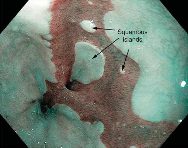
Figure 2.
Endoscopic photograph of Barrett's esophagus using narrow band imaging. The metaplastic columnar (Barrett's) epithelium is dark, and the squamous epithelium is light. Notice the squamous islands, which presumably develop as a consequence of biopsy sampling of metaplastic epithelium during endoscopic surveillance.

