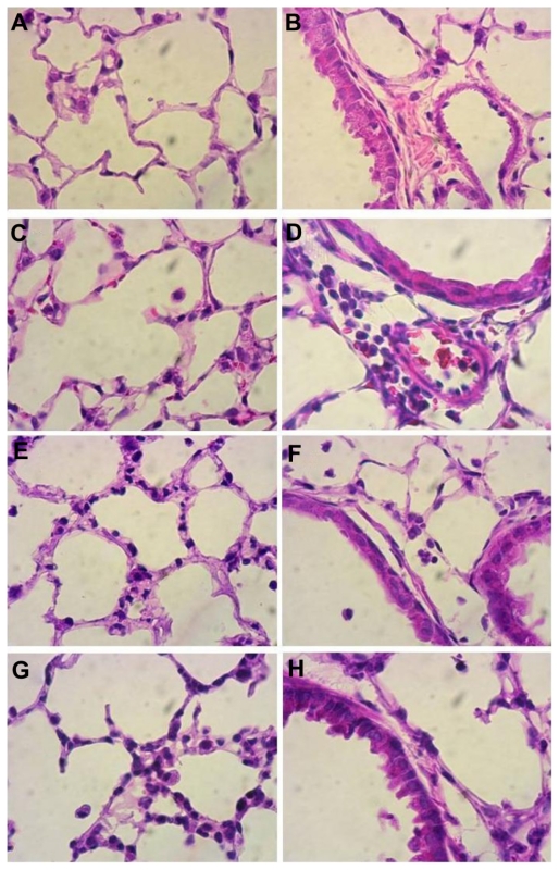
Figure 2.
Histopathology of lung sections. Lungs from mice treated intranasally with 80 μg lipopolysaccharide and/or K90/RGDSK10 rosette nanotubes (5%, 12.5 μL intravenously) and saline controls were evaluated using sections stained with hematoxylin and eosin. Control lung tissue showed no inflammation and normal lung architecture (A, B), whereas mice treated with K90/RGDSK10 rosette nanotubes (C, D), lipopolysaccharide (E, F), and lipopolysaccharide + K90/RGDSK10 rosette nanotubes (G, H) showed septal neutrophilic infiltration, leukocytes adhering to the blood vessel wall, perivascular infiltration of leukocytes, and peribronchiolar accumulation of neutrophils at the 12-hour time point.

