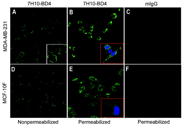
Figure 3.
Immunofluorescence localization of the ATP synthase α-subunit on the surface of breast cancer cells by confocal microscopy. Cells were stained with an α-ATP synthase antibody, followed by secondary antibody, as detailed in Materials and Methods. A and D, Non-permeabilized MDA-MB-231 and MCF-10F immunostained with a murine mAb specific for the ATP synthase α-subunit(600×). B and E, Image obtained from MDA-MB-231 and MCF-10F cells permeabilized with ethanol (100%). C and F, Control experiments for antibody specificity using isotypic purified mouse IgG in cells(600×).

