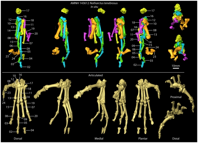
Figure 4. Pedal elements after preparation.
After physical preparation of the foot was completed, all bones were scanned with microCT at resolutions ranging from 0.013–0.031 millimeter voxels. High resolution surface files were created from these images. One set of images was overlaid on the original CT scan shown in Fig. 3 to allow easier viewing and study of the in situ elements (top row). Another set of 3D surface images were articulated in a “closest packed” arrangement to get a better sense of what the foot looked like in the living animal (bottom row).

