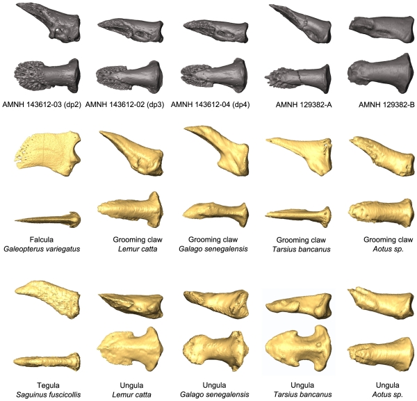
Figure 9. Comparison of fossil and extant distal phalanges.
MicroCT images of distal phalanges are displayed in two views: lateral (above) and dorsal (below). Fossil unguals are shown in comparison to extant specimens that bear different unguis forms: falculae (claws), grooming claws, tegulae, and ungulae (nails).

