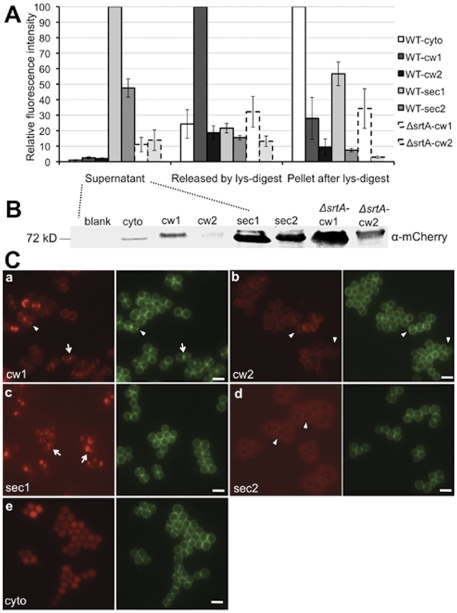
Figure 2. Monitoring mCh-hybrids.
A. Fluorescence intensity comparison of mCh-hybrids from different cell fractions. WT-cyto, SA113 (pCXmCh-cyto); WT-cw1 or 2, SA113 (pCXmCh-cw1) or (pCXmCh-cw2); WT-sec1 or 2, SA113 (pCXmCh-sec1) or (pCXmCh-sec2); ΔsrtA-cw1 or 2, SA113 ΔsrtA (pCXmCh-cw1) or (pCXmCh-cw2); lys, lysostaphin. B. Western blotting of mCh-hybrid proteins in the culture supernatant of protein A deficient mutant SA113 Δspa. Blank, SA113 Δspa without plasmid; cyto, SA113 Δspa (pCXmCh-cyto); cw1 or 2, SA113 Δspa (pCXmCh-cw1) or (pCXmCh-cw2); sec1 or 2, SA113 Δspa (pCXmCh-sec1) or (pCXmCh-sec2); ΔsrtA-cw1 or 2, SA113 ΔspaΔsrtA (pCXmCh-cw1) or (pCXmCh-cw2). C. Subcellular localization of mCh-hybrid proteins in SA113. a. pCXmCh-cw1; b. pCXmCh-cw2; c. pCXmCh-sec1; d. pCXmCh-sec2; e. pCXmCh-cyto. Arrowheads in a and b, fluorescence localized at the cross wall in a, but absent from the cross wall in b; arrows in a and c, RF foci close to the initial sites of the cross walls; arrowheads in d, halo-like RF distribution absent from the cross wall. Images a, c, and e were taken after one hour of xylose induction; images b and d were taken after two hours of induction. Green: Van-FL staining (cell wall); scale bar, 2 µm.

