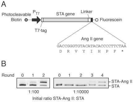
Figure 3. Construction and enrichment of the streptavidin-fused angiotensin II (STA-Ang II) gene in multiple rounds of DNA-display selection on hAT1R/CHO-K1 cells.
(A) A schematic representation of the DNA template for in vitro transcription/translation. DNA was labeled during PCR with photocleavable biotin [22] at the upstream ends and with fluorescein at the downstream ends, using labeled primers. The translated open reading frame consists of sequences for a T7·tag, streptavidin (STA), a peptide linker, and Ang II gene. The 5′-UTR fragment contains T7 promoter. (B) Reaction mixtures containing 1∶100 or 1∶10,000 molar ratio of STA-Ang II: STA genes were emulsified. The DNA after each round of selection was PCR-amplified with a fluorescein-labeled primer and analyzed by 15% PAGE with an imaging analyzer.

