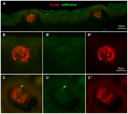
Figure 5. Double immunolabeling for PLCβ2 (red) and βgal (green) in the palate and fungiform taste fields where A2BR-βgal expression is weak or absent shows specificity of the anti-gp secondary antiserum as well as lack of staining for βgal.
Panels A and B show palatal taste buds; the lack of green staining (A; B, B′) shows both the absence of βgal immunoreactivity and the lack of cross reactivity of the anti-gp secondary antiserum with the rb PLCβ2 primary antiserum plainly visualized with the red anti-rb secondary antiserum (B, B″). Panel C shows a fungiform taste bud where only one (green arrow) of the PLCβ2-positive cells (red, C, C″)) shows faint reaction for βgal (green, C, C′). The lack of green label in the other strongly positive red cells again demonstrates specificity of the anti-gp (green) antiserum. B′, B″ and C′, C″ show color separation images of panels B and C respectively. Scale bar in B″ also applies to C.

