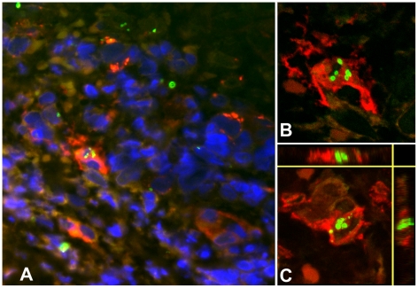
Figure 3. Multiple intracellular O. tsutsugamushi within antigen presenting cells (APCs) in the superficial dermis.
APCs are characterised by MHC class II receptor (HLADR) positivity and were associated with intracellular O. tsutsugamushi in admission samples of an eschar from a patient with acute scrub typhus. Panel A: HLADR-positive cells in red, O. tsutsugamushi in green. Panels B and C: Laser Scanning Micrographs. Panel B depicts the same infected cell as in Panel A as a 0.3 µm thin section. Panel C shows a 3D stack projection (of 0.3 µm thin sections) of intracytosolic O. tsutsugamushi (in green), with small, high-density granules with high refractive index, staining positively for O. tsutsugamushi antigen. Patient TM2193, magnification ×400, LSM inserts ×1000, double-immunolabeling: HLADR in red, O. tsutsugamushi in green and DAPI nuclear counterstain in blue.

