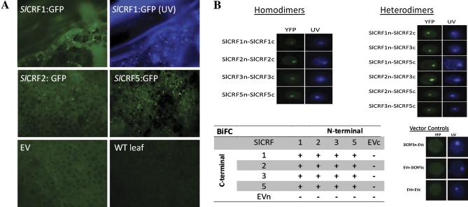
Fig. 4.
SlCRF protein localization and protein–protein interactions. (A) Cellular localization of SlCRF1, SlCRF2, and SlCRF5 in tobacco leaves transiently transformed with 35S:SlCRF:GFP vectors via Agrobacterium infiltration. Representative examples of GFP expression from tagged SlCRF proteins indicate a strong nuclear localization in regions of transformed leaves visualized under UV light using a GFP wavelength filter (panels labelled SlCRF:GFP).The panel labelled SlCRF1:GFP (UV) is the same sample as SlCRF1:GFP shown without the GFP filter in the presence of Hoechst 33342 dye denoting the nucleus. EV denotes an empty vector control and WT leaf denotes an untransformed sample. (B) SlCRF proteins (SlCRF1, SlCRF2, SlCRF3, and SlCRF5) were analysed for potential homo- and heterodimerization using BiFC. Representative examples of positive SlCRF dimerizations are shown both under UV light in the presence of Hoechst 33342 dye denoting the nucleus and using a YFP wavelength filter to visualize BiFC interaction. Additionally, representative examples of empty vector (EV) controls for both N- and C-terminal BiFC vectors (EVn and EVc) are shown. A table of SlCRF interactions is shown, with (+) as positive and (–) for non-interactions.

