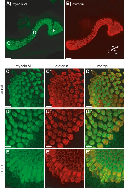
FIG. 6.
Otoferlin is present in all AP hair cells. A 10× image of the AP that has been stained for myosin VI (A) and otoferlin (B). The orientation of the papilla is indicated by the inset in (B), where C caudal, R rostral, M medial, and L lateral. C 63× images of the caudal region of the AP showing labeling of myosin VI (C), otoferlin (C′) and their merged image (C″). D, D′, D″ as in (C), but from the medial portion of AP. E, E′, E″ as in (C) but from the rostral portion of the AP. The approximate location for these higher magnification images are indicated in (A). Scale bars: 96 μm (A, B); 14 μm (C–E).

