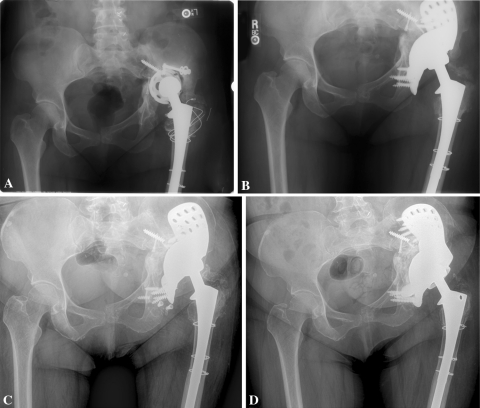
Fig. 2A–D.
(A) A preoperative AP pelvic radiograph demonstrates a failed acetabular component with pelvic discontinuity. (B) An immediate postoperative AP pelvic radiograph demonstrates a well-fixed triflange component. (C) The triflange component failed due to aseptic loosening at 11 years postoperatively. (D) The revision triflange component is well fixed at 6 months.

