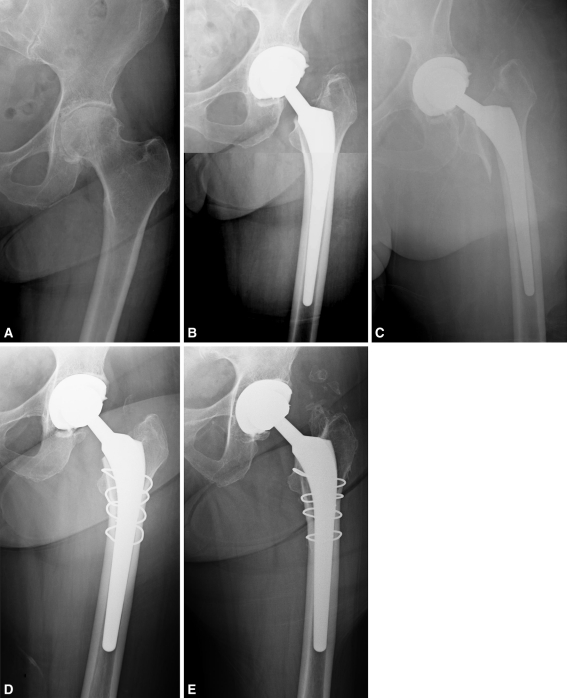
Fig. 4A–E.
(A) A preoperative radiograph of the left hip of a 79-year-old female patient who presented with severe pain and discomfort secondary to osteoarthritis shows severe joint space narrowing, sclerosis, and osteophyte and cyst formation. (B) An immediate postoperative radiograph shows treatment of cementless primary left THA with an 11- by 160-mm standard-length taper stem with lateralized offset and a 36-mm cobalt-chromium head with +6-mm neck articulated against highly crosslinked polyethylene. (C) At 2 weeks postoperative, the patient presented emergently with severe pain. A radiograph reveals a periprosthetic femoral fracture. (D) The patient’s femoral component was revised to a 14- by 175-mm standard-length tapered stem with lateralized offset, and the bone was secured with five cerclage cables. (E) A radiograph at 3 years after revision shows well-fixed components in satisfactory position and alignment. The patient had a HHS of 94 at latest followup.

