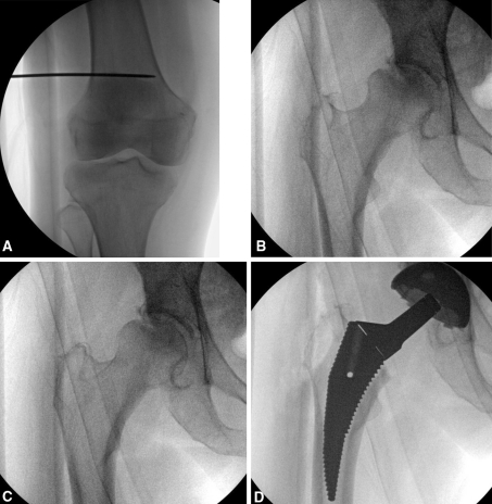
Fig. 2A–D.
(A) AP view of the knee with a unicortical Kirschner wire placed. (B) AP of the hip corresponding to AP view of the knee in A. (C) Internally rotated view of the hip until a single profile of the greater trochanter; degree of rotation designated the anteversion angle of the hip. (D) True AP of the rasp, noted by a perfect circle for the hole in the rasp, with rotation measured from the Kirschner wire designating the stem anteversion.

