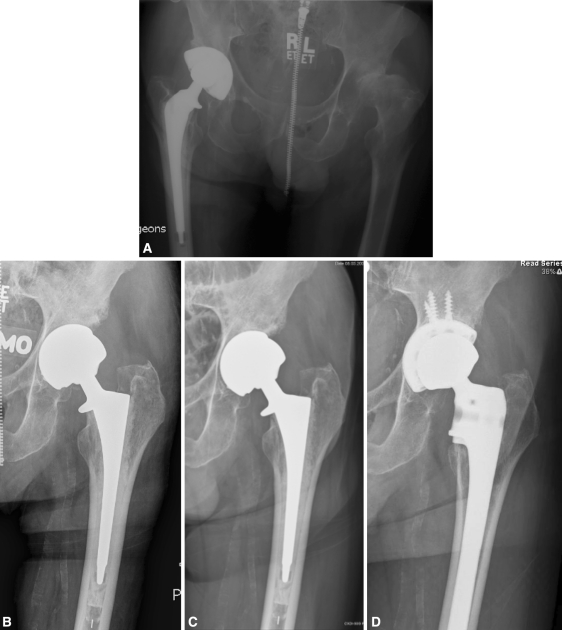
Fig. 3A–D.
Radiographs illustrate the case of a 68-year-old man who underwent LD-MOM THA of the left hip 17 years after THA of the right hip. (A) A preoperative radiograph shows avascular necrosis and superolateral subluxation of the left hip. (B) The initial 2-month radiograph of the Magnum™ THA shows incomplete seating of the shell against the medial wall. (C) The patient lived with pain for 12 months before revision THA. Note inferior osteolysis and progressive loosening of the cup. (D) At revision, the cemented stem was also loose. Workup for infection was unremarkable.

