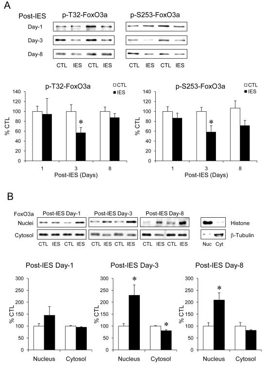
Figure 1.
IES-induced activation of FoxO3a in the cerebral cortex. The cerebral cortex from control (CTL) and IES-treated mice were dissected on post-IES day-1 (n=6/group), day-3 (n=14/group), and day-8 (n=8/group). (A) Protein homogenates were immunobloted for phospho-T32-FoxO3a, phospho-S253-FoxO3a, and total FoxO3a. (B) Nuclear and cytosolic proteins were immunobloted separately for total FoxO3a. Histone and β-tubulin were immunobloted as quality control of nuclear/cytosolic extraction. Data is expressed as % control (no IES). Mean ± SEM, *p<0.05 in Student’s t-test when IES-treated mice were compared to control mice.

