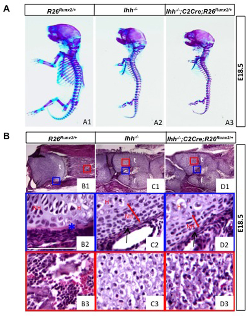
Fig. 5.

Morphological analyses of Ihh−/− embryos expressing Runx2. (A) Whole-mount skeletal staining. (B) H&E staining of longitudinal sections of the tibia. Proximal ends to the left. Boxed areas in B1-D1 shown at a higher magnification in B2-D2 and B3-D3. Scale bar: 100 µm. t: tibia; f: fibula; NH: nonhypertrophic chondrocytes; H: hypertrophic chondrocytes. Arrows denote hypoplastic perichondrium. Asterisk denotes bone collar.
