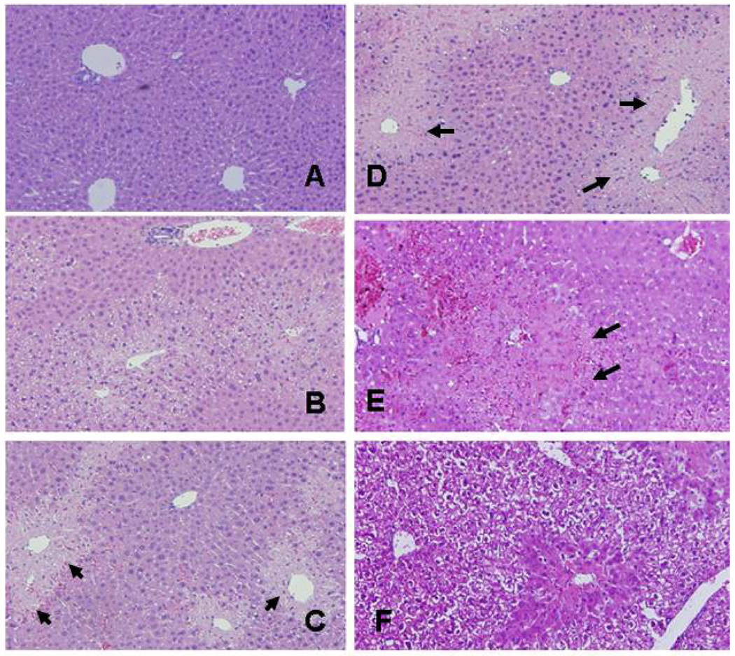
Figure 2. Effect of APAP toxicity on liver histology.
Mice were treated with APAP (200 mg/kg IP) and sacrificed at 2, 4, 8 or 24 h (panels B-E, respectively). Some mice received saline (panel A). Other mice received APAP+NAC (panel F). Arrows indicate areas of necrosis. At 2 h (B), there was centrilobular pallor in the hepatocytes surrounding the central vein and the nuclei were shrunken in some cells. By 4 h (C), the regions of necrosis were defined and progressed in extent by 8 h (D). By 24 h (E), the central veins were collapsed and filled with cellular debris and frank hemorrhage was present. The progression of necrosis was halted in the mice treated with APAP+NAC (F).

