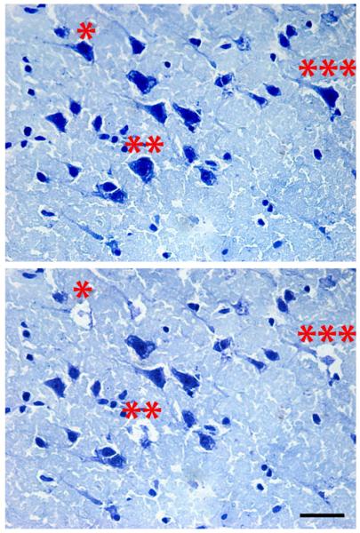
Fig. 2.
Photomicrograph of pyramidal cells from Layer V from ventral bank of the principal sulcus of the DLPFC. (Top panel) Representative pyramidal neurons selected for microdissection based on morphology and cortical location. (Bottom panel) Same tissue section shown in A illustrating the areas of microdissection. Note the specificity of the dissection and the minimal disruption of surrounding neuropil. Asterisks indicate cells dissected from a representative section (scale bar=500 μm).

