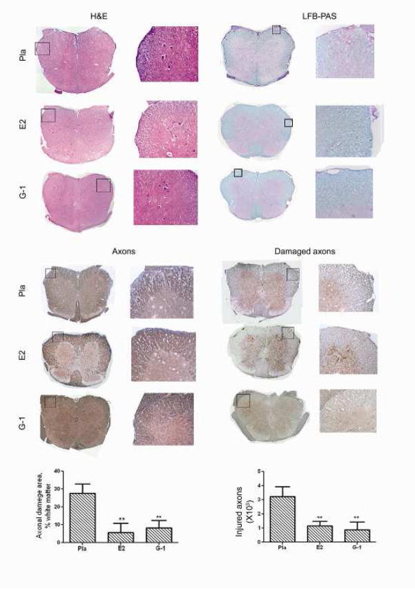
Fig. (4). G-1 treatment reduced CNS infiltration (H&E), demyelination (LFB-PAS), axonal loss (NFLs) and ongoing axonal damage (dephosphorylated NFLs).
The mice from the clinical experiment shown in Fig. 2a were euthanized at the end of the experiment and spinal cords from ≥3 mice from each group were dissected for histology. Immune cell infiltration and demyelination of CNS were examined with H&E and LFB-PAS staining. Total and damaged axons were examined with immunohistological staining for neurofilaments (NFLs) or dephosphorylated NFLs.

