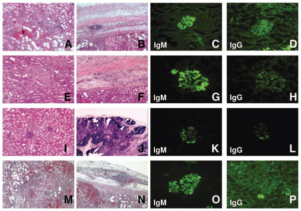
FIGURE 3.
Group 2 and 3: Graft histology. Histology of B256 (group 2, animal no. 1) on POD15 revealed the TK graft was injured, but not rejected (A, B). Immunohistochemistry showed frequent glomerular anti-non-Gal IgM and scattered anti-non-Gal IgG in few glomeruli (C, D). Histology of B257 (group 2, animal no. 2) after TK transplantation with a regimen of anti CD3-IT T-cell depletion (hematoxylin-eosin staining): the TK graft had mild hemorrhage and multiple microthrombi on POD15 (E, F). Immunohistochemistry showed IgM deposition in most glomeruli (G) with weak IgG deposition in some of the glomeruli (H). The transplanted TK graft was accepted on day 8 after Re–TK transplantation (I, J). Immunohistochemistry showed deposition of IgM in many glomeruli (K) but few IgG deposits (L). Histology of B272 (group 3, animal no. 2) after TK transplantation with a regimen of LoCD2 T-cell depletion (hematoxylin-eosin staining). Cellular rejection and advanced antibody mediated rejection developed (M). Hemorrhage was also seen in the thymic tissue under the renal capsule (N). Immunohistochemistry findings demonstrated both anti-non-Gal IgM and IgG deposition in glomeruli (O, P).

