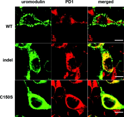
Figure 6.
Cellular distribution of wild-type (WT) and mutant uromodulin proteins expressed in HEK293 cells, 48 hours post-transfection. Cells transfected with WT, indel, or C150S UMOD were costained with anti-uromodulin (left panels, green) and anti-PD1 (middle panels, red) antibodies. (right panels) The merged images clearly show colocalization of uromodulin and the ER-resident protein PD1, in both indel- and C150S-expressing cells. Bar, 5 μm.

