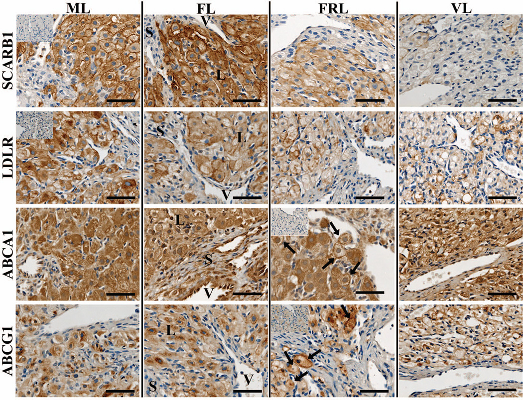
Figure 2. Localization of lipoprotein receptors and ABC transporters in the rhesus macaque CL throughout the period of spontaneous functional regression.
Representative photomicrographs of SCARB1, LDLR, ABCA1, and ABCG1 from the mid-late (ML), functional late (FL), functionally-regressed late (FRL), and very-late (VL) luteal stages are shown. The insets in the upper left corner of the ML samples for SCARB1 and LDLR, as well as in the FRL samples for ABCA1 and ABCG1, are sections that were processed with primary antibody preabsorbed with immunizing peptide. The approximate locations of various cell types are indicated in the FL panel for each protein and include: large luteal cells (L); small luteal or stromal cells (S); and blood vessels (V). The scale bar in the lower right hand corner of each image is 50 µm. Arrows indicate the appearance of plasma membrane localization of ABCA1 and ABCG1 in FRL stage CL.

