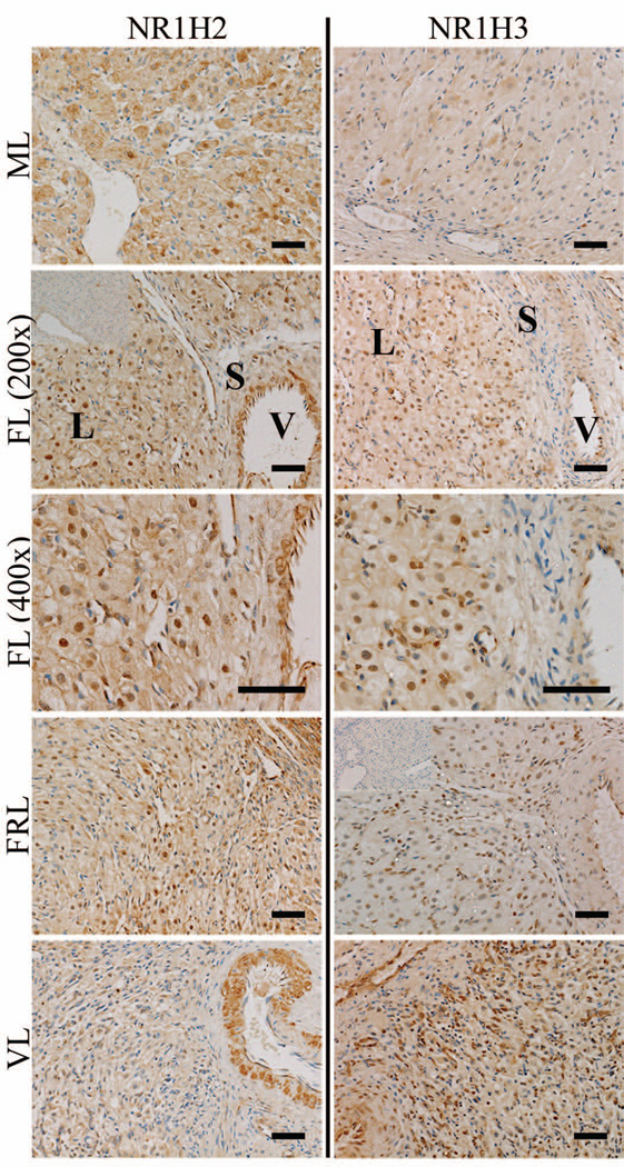
Figure 6. Localization of NR1H2 and NR1H3 expression in the rhesus macaque CL throughout the period of spontaneous functional regression.
Representative photomicrographs of NR1H2 and NR1H3 from the mid-late (ML), functional late (FL), functionally-regressed late (FRL), and very-late (VL) stages are shown. The insets in the upper left corner of the FL 200× panel for NR1H2, and FRL panel for NR1H3, are control sections incubated with primary antibody preabsorbed with its immunizing peptide (NR1H2), or where the primary antibody was excluded (NR1H3). The approximate locations of various cell types are indicated in the FL panel for each protein and include: large luteal cells (L); small luteal or stromal cells (S); and blood vessels (V). The scale bar in the lower right hand corner of each image is 50 µm.

