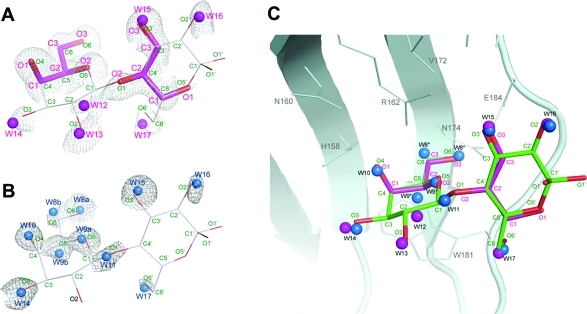
Figure 2.
Carbohydrate-binding site of Gal3C. The 2Fo – Fc electron density maps, contoured at 1.0σ, are shown for the binding sites of glycerol-bound Gal3C and apo-Gal3C, superimposed on the structure of lactose (stick model) in lactose-bound Gal3C. (A) Electron density for glycerol-bound Gal3C. Glycerol and water molecules in the glycerol compex are shown as magenta sticks and spheres, respectively. To illustrate the molecular mimicry of lactose by glycerol, lactose is shown as thin sticks. The lactose and glycerol atom annotations are colored green and magenta, respectively. (B) Electron density for water molecules in apo-Gal3C. Water molecules are shown as blue spheres. Labels a and b are used to denote alternate positions of water molecules W8 and W9. As in panel A, lactose is shown as thin sticks as an aid to interpretation. (C) Superposition of the lactose-bound Gal3C, glycerol-bound Gal3C, and apo-Gal3C structures, demonstrating the common oxygen recognition motif of the binding site. The oxygens of lactose (green) and glycerol (magenta) occupy the same positions as water molecules found in apo (blue spheres) and glycerol-bound (magenta spheres) Gal3C.

