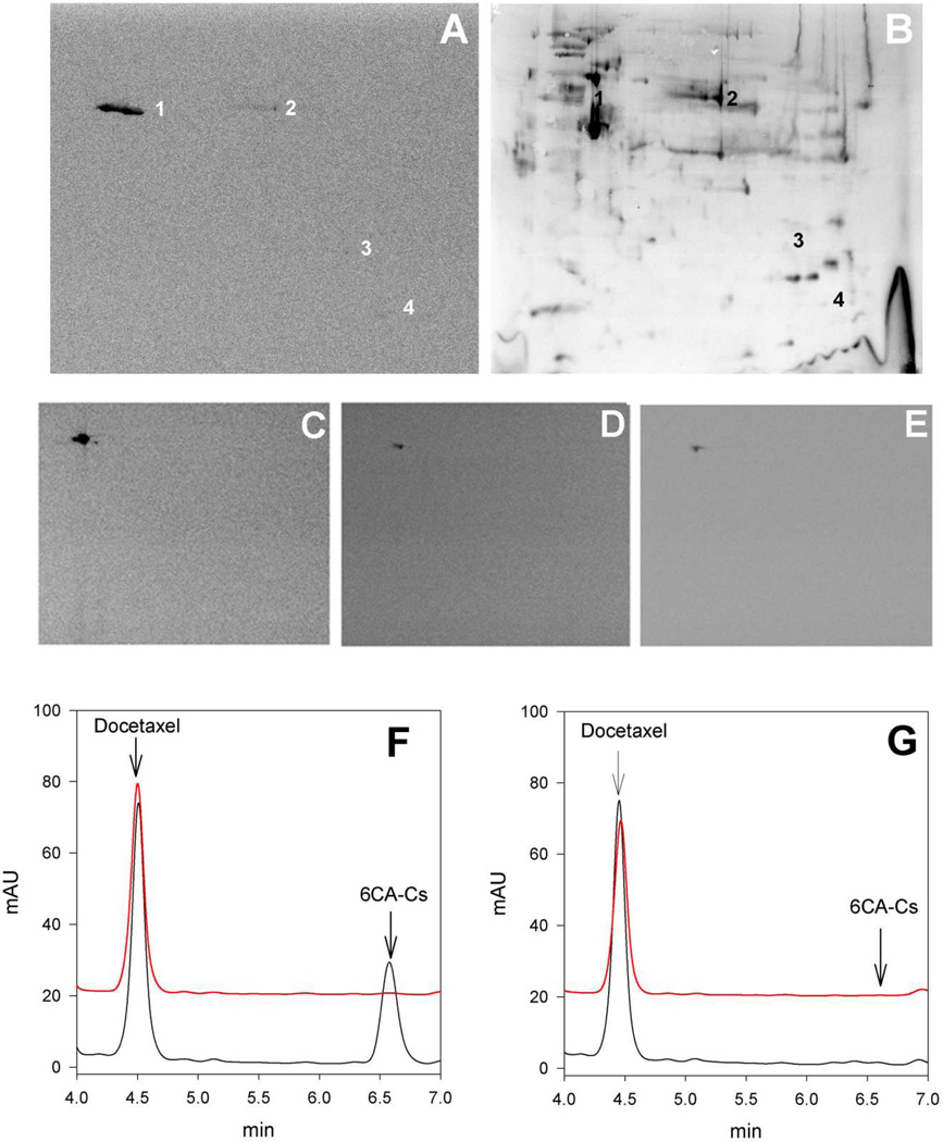
Figure 3.
Upper Panel Identification of [14C]8Ac-Cs protein cell targets. Two dimensional gel electrophoresis of proteins extracted from A549 cells incubated with 2.5 µM [14C]8Ac-Cs for 24 h. (A) Autoradiogram of the PDVF membrane after protein transfer. (B) The silver-stained 2D replica gel obtained with the protein extracts from treated cells and the spots corresponding to radiolabeled proteins are indicated (1 to 4). Protein identification was performed by in-gel trypsin digestion followed by database search with MALDI-TOF-MS data. Panels C,D,E.-[14C]8Ac-Cs cell target is conserved in different cell lines. 1A9 cells incubated with 2.5 µM (C) or 300 nM [14C]8Ac-Cs (D) and A549 cells incubated with 300 nM [14C]8Ac-Cs (E). Lower Panel. HPLC analysis of 6CA-Cs extracted from pellets (red) and supernatants (black) after incubation without (F) and with (G) stabilized MTs.

