Figure 5.
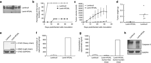
Tumor-suppressing activity of lenti-HF2AL in vivo. HCT116 cells (5 × 10⁵) infected with either lenti-HF2AL or lenti-null (MOI = 25) were injected into subcutaneous tissue of the right dorsal flank of 6-week-old female nude mice (n = 6). On day 32, the mice were killed, and serum and tumor tissues were obtained and analyzed. (a) Tumor formation in the mice at day 32 (b) or throughout the whole time course of the experiment is shown. (c) Tumor size and (d) tumor weight were decreased by lenti-HF2AL infection. (e) Expression of zaptuximab in the tumor tissues was detected by western blot analysis and (f) enzyme-linked immunosorbent assay (ELISA). (g) Quantitative expression of zaptuximab in the sera samples were detected by ELISA. (h) Activation of caspase-3 was detected in the tumor tissue. A total of 100 µg of protein from the lysates prepared from tumor tissues of the above mice was separated by SDS-PAGE. Activation of caspase-3 was examined using immunoblotting with the corresponding antibodies. GAPDH was used as the loading control. GAPDH, glyceraldehyde-3-phosphate dehydrogenase; MOI, multiplicity of infection; SDS-PAGE, sodium dodecyl sulfate-polyacrylamide gel electrophoresis.

