Figure 1.
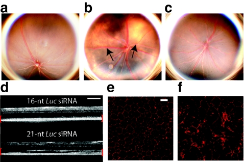
21-Nucleotide (nt) short-interfering RNAs (siRNAs) induce retinal degeneration. (a) Representative fundus photographs of mice 7 days after treatment with intravitreous phosphate-buffered saline (PBS) control and (b) 21-nt-Luc-siRNA demonstrating large hypopigmented areas indicative of retinal pigment epithelium (RPE) loss (black arrows, n = 24 per group in four independent experiments, P < 0.0001 by Fisher's exact test). (c) Administration of 16-nt-Luc-siRNA did not induce retinal degeneration (n = 24 in four independent experiments, P < 0.0001). (d) In vivo retinal imaging with optical coherence tomography revealed marked disruption of the outer retina and RPE (enclosed in red brackets) with 21-nt Luc siRNA (n = 6 in two independent experiments). Zonula occludens-1 (ZO-1) immunofluorescence (red) of RPE flat mounts 1 week after 16-nt-Luc-siRNA (e) showed uniformly arranged hexagonal cells, but 21-nt-Luc-siRNA treatment (f) resulted in significant disruption of this junctional pattern with large variations in cellular morphology (n = 4 in two independent experiments). Bars (d) = 200 µm; Bars (e, f) = 20 µm.

